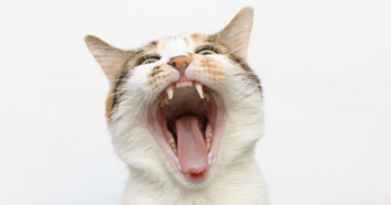
If your beloved feline is over five years old, there is a good chance he or she has a painful feline odontoclastic resorptive lesion (FORL), one of the more common oral abnormalities we see in veterinary practice. These dental resorptions are also sometimes called cat cavities, caries, cervical neck lesions, external or internal root resorptions, and cervical line erosions. FORLs are usually found on the outside surface of the tooth where the gum meets the tooth surface. Although the premolars of the lower jaw are most commonly affected, lesions can be found on any tooth in any location.
Unlike cavities in humans, which are the result of bacterial enzymes and acids digesting the teeth, the cause of FORLs is not completely understood. Cells known as odontoclasts are found in the defects causing the tooth structure to dissolve but the trigger of this reaction is not clear. A reaction to plaque on the teeth seems to be the major factor (another reason daily brushing is beneficial for all felines). Research suggests a correlation between problems with calcium metabolism, chronic calicivirus infections, or an autoimmune response.
So how do you know if your cat is in the majority and has a resorptive lesion?
Cats affected with FORLs may show increased salivation, oral bleeding, or have difficulty chewing. They may drop food from their mouths while eating or only appear to eat on one side of their mouth. Unfortunately most cat parents notice no obvious signs of the dental disease.
If your vet suspects your cat may have a FORL, they may use a cotton-tipped applicator to press against the suspected lesion. This causes pain when the FORL is touched and the cat's jaw actually spasms. The trembling is not subtle and is often alarming to the pet parent. While this is clearly not pleasant for the patient, this is the best way I know to demonstrate to the pet parent the pain their cat is in and likely has been experiencing for some time. In some cases, the FORL will be covered with inflamed gum tissue and is only detected while the patient is under anesthesia having a dental cleaning performed.
FORLs vary in severity and so does their treatment. Therapy of mild defects (stage one) usually involves thorough cleaning and smoothing the defect with a dental drill. Slightly more advanced lesions (stage two) may benefit from restorative treatment.
X-rays are necessary when the lesion progresses to stage three. Either a root canal or extraction is typically needed at this stage. There may even be abscesses associated with this advanced lesion. The most advanced FORLs, stage four, are ones in which the gum has actually grown over the remnants of the tooth, perhaps to protect the painful fragments remaining. Treatment requires extraction of the root fragments if they appear inflamed or painful to the patient. This is the most difficult stage to treat, as the remaining tooth is often prone to fracturing into numerous pieces. You an imagine that hunting for barely visible tooth root fragments through a small hole in your cat's gum is not easy, but your veterinarian is trained to do this. Still, it is far easier on everyone involved (especially the cat) to catch and treat FORLs at an early stage.
To be on FORL lookout at home, examine your cat’s mouth for FORLs monthly. Take a Q-tip and gently place it against the area where the tooth meets the gum. Examine the entire mouth if possible, but if your feline is displeased about having a q-tip in his mouth, focus on the lower jaw teeth first. If there is pain or bleeding a trip to the veterinarian is in order. Your best friend will certainly thank you (as will your veterinarian - for being an educated advocate for your cat)!